Please use this identifier to cite or link to this item:
https://doi.org/10.21256/zhaw-3590Full metadata record
| DC Field | Value | Language |
|---|---|---|
| dc.contributor.author | Bono, Epifania | - |
| dc.contributor.author | Mathes, Stephanie | - |
| dc.contributor.author | Rimann, Markus | - |
| dc.date.accessioned | 2018-04-20T12:37:42Z | - |
| dc.date.available | 2018-04-20T12:37:42Z | - |
| dc.date.issued | 2017 | - |
| dc.identifier.uri | https://digitalcollection.zhaw.ch/handle/11475/5440 | - |
| dc.description.abstract | Introduction: Drug-induced liver injury is the leading cause of acute liver failure and post-market drug withdrawals. In vivo animal studies cannot be totally translated to humans; therefore, there’s huge demand of novel in vitro human models. 3D culture conditions would increase models longevity while bioprinting technology is expected to improve their functionality by the in vivo-like cell spatial patterning. Aim: The present project aims at the establishment and characterization of printed liver tissue-like models as co-culture of human cells such as hepatocytes, stellate and endothelial cells in order to reproduce a functional liver sinusoid. 3D equivalents were printed to assess hepatic cells printability (printing test), while high-density hepatocytes models were manually produced and characterized in order to simulate in-vivo cellular density conditions and define experimental parameters for bioprinting process. Materials and methods: - Printing test: HepG2 cells were mixed 6*106 cells/ml with bioink, PEG-based ink produced in house. Matrigel solution was added to further improve cell viability during the printing process. Cell mixture was printed by direct dispensing in a spiral pattern and polymerized at 365nm wavelength. Models were analysed up to 7 days for cell proliferation, viability and morphology. - High-density 3D models: High-density HepG2 discs and drop models were manually produced mixing 2:1 HepG2 “paste” with ink (supplemented with matrigel). Models were polymerized by exposure to 365nm wavelength for few seconds and cultivated up to 28 days. The equivalents were analysed for cell viability and albumin secretion as well as processed for histological analyses (cell proliferation, tissue-like intercellular tight junctions and lipid storage investigation). Results and discussion: - Bioprinting as well as ink is suitable for HepG2 viability and proliferation up to 7 days (15.84 ± 1.46 fold change increase day 7 vs day1). Printed HepG2 are homogenously distributed and round-shaped, forming agglomerates that increase in size over time. - HepG2-ink-matrigel equivalents are long-term stable characterized by high and constant cell viability up to 28 days. The models resemble native liver with respect to the high cell density and due to the ink supplementation can easily be printed into tissue-like models with exact cell patterning and defined structure. Models are currently under investigation for tissue functionality and morphology and will be subsequently printed. Conclusions: Bioprinting shows high potential for the manufacture of high-density liver tissue-like models. The printing of different cell types (hepatocytes, stellate and endothelial cells) will allow producing organotypical 3D liver equivalents. | de_CH |
| dc.language.iso | en | de_CH |
| dc.publisher | ZHAW Zürcher Hochschule für Angewandte Wissenschaften | de_CH |
| dc.rights | Licence according to publishing contract | de_CH |
| dc.subject | Bioprinting | de_CH |
| dc.subject | 3D hepatic model | de_CH |
| dc.subject.ddc | 571: Physiologie und verwandte Themen | de_CH |
| dc.subject.ddc | 572: Biochemie | de_CH |
| dc.subject.ddc | 660.6: Biotechnologie | de_CH |
| dc.title | Evaluation of 3D hepatic tissue models for bioprinting | de_CH |
| dc.type | Konferenz: Poster | de_CH |
| dcterms.type | Text | de_CH |
| zhaw.departement | Life Sciences und Facility Management | de_CH |
| zhaw.organisationalunit | Institut für Chemie und Biotechnologie (ICBT) | de_CH |
| dc.identifier.doi | 10.21256/zhaw-3590 | - |
| zhaw.conference.details | TERMIS-EU 2017 Conference, Davos, 26-30 June 2017 | de_CH |
| zhaw.funding.eu | No | de_CH |
| zhaw.originated.zhaw | Yes | de_CH |
| zhaw.publication.status | publishedVersion | de_CH |
| zhaw.publication.review | Not specified | de_CH |
| zhaw.webfeed | 3D Gewebe und Biofabrikation | de_CH |
| Appears in collections: | Publikationen Life Sciences und Facility Management | |
Files in This Item:
| File | Description | Size | Format | |
|---|---|---|---|---|
| TERMIS Poster 2017_liver printing_final.pdf | 326.99 kB | Adobe PDF | 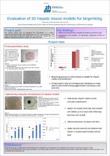 View/Open |
Show simple item record
Bono, E., Mathes, S., & Rimann, M. (2017). Evaluation of 3D hepatic tissue models for bioprinting. TERMIS-EU 2017 Conference, Davos, 26-30 June 2017. https://doi.org/10.21256/zhaw-3590
Bono, E., Mathes, S. and Rimann, M. (2017) ‘Evaluation of 3D hepatic tissue models for bioprinting’, in TERMIS-EU 2017 Conference, Davos, 26-30 June 2017. ZHAW Zürcher Hochschule für Angewandte Wissenschaften. Available at: https://doi.org/10.21256/zhaw-3590.
E. Bono, S. Mathes, and M. Rimann, “Evaluation of 3D hepatic tissue models for bioprinting,” in TERMIS-EU 2017 Conference, Davos, 26-30 June 2017, 2017. doi: 10.21256/zhaw-3590.
BONO, Epifania, Stephanie MATHES und Markus RIMANN, 2017. Evaluation of 3D hepatic tissue models for bioprinting. In: TERMIS-EU 2017 Conference, Davos, 26-30 June 2017. Conference poster. ZHAW Zürcher Hochschule für Angewandte Wissenschaften. 2017
Bono, Epifania, Stephanie Mathes, and Markus Rimann. 2017. “Evaluation of 3D Hepatic Tissue Models for Bioprinting.” Conference poster. In TERMIS-EU 2017 Conference, Davos, 26-30 June 2017. ZHAW Zürcher Hochschule für Angewandte Wissenschaften. https://doi.org/10.21256/zhaw-3590.
Bono, Epifania, et al. “Evaluation of 3D Hepatic Tissue Models for Bioprinting.” TERMIS-EU 2017 Conference, Davos, 26-30 June 2017, ZHAW Zürcher Hochschule für Angewandte Wissenschaften, 2017, https://doi.org/10.21256/zhaw-3590.
Items in DSpace are protected by copyright, with all rights reserved, unless otherwise indicated.