Please use this identifier to cite or link to this item:
https://doi.org/10.21256/zhaw-1648Full metadata record
| DC Field | Value | Language |
|---|---|---|
| dc.contributor.author | Crawford, Rebecca | - |
| dc.contributor.author | Volken, Thomas | - |
| dc.contributor.author | Valentin, Stephanie | - |
| dc.contributor.author | Melloh, Markus | - |
| dc.contributor.author | Elliott, James Matthew | - |
| dc.date.accessioned | 2018-02-07T15:04:17Z | - |
| dc.date.available | 2018-02-07T15:04:17Z | - |
| dc.date.issued | 2016-08-05 | - |
| dc.identifier.issn | 2397-1789 | de_CH |
| dc.identifier.uri | https://digitalcollection.zhaw.ch/handle/11475/2675 | - |
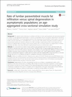
| dc.description.abstract | Background: The spinal column including its vertebrae and disks has been well examined and extensively reported in relation to age-aggregated degeneration. In contrast, paravertebral muscles are poorly represented in describing normative degeneration. Increasing evidence points to the importance of paravertebral muscle quality in low back health, and their potential as a modifiable factor in low back pain (LBP). Studies examining normative decline of paravertebral muscles are needed to advance the field’s etiological understanding. With a novel approach and based on published data, we establish and compare decline rates of imaging features for degeneration of lumbar vertebrae and disks, versus fatty infiltration in paravertebral muscles in asymptomatic adults.Methods: Our cross-sectional simulation study examined age-aggregated data from three published studies who reported on asymptomatic adults spanning 18–60 years. Prevalence rates of imaging degenerative features of the spinal column were examined via logistic regression and compared with percentage fatty infiltration in erector spinae, multifidus and psoas using synthetic data and Monte Carlo simulation with 10,000 endpoint-specific regression iterations. General linear regression models were employed to estimate marginal effects of age reported as a one-year change rate (with 95 % confidence intervals) for comparisons between all reported spinal features. Results: Declines in multifidus (0.24 & 0.11 %/year), erector spinae (0.13 & 0.07 %/year), and psoas (0.04 %/year) occur at similarly slow rates to disk protrusion (0.25 %/year), annular fissure (0.15 %/year), and spondylolisthesis (0.29 %/year). Multifidus showed a trend for faster decline than erector spinae, particularly in men. Of the features examined, disk signal loss declined fastest, and psoas muscle the slowest. Conclusions: Degeneration of lumbar paravertebral muscles occurs slowly in asymptomatic adults, with a tendency to be most pronounced in multifidus. Rate of decline of spinal structures represents a novel variable that warrants inclusion as a known feature of the expected degenerative cascade, and to provide a basis for comparison to diseases of the spine in research and clinical practice. Concurrent examination of spinal features using advanced imaging to improve muscle analysis would be a strong addition to the field. | de_CH |
| dc.language.iso | en | de_CH |
| dc.publisher | BioMed Central | de_CH |
| dc.relation.ispartof | Scoliosis and Spinal Disorders | de_CH |
| dc.rights | http://creativecommons.org/licenses/by/4.0/ | de_CH |
| dc.subject | Lumbar spine | de_CH |
| dc.subject | Paravertebral muscles | de_CH |
| dc.subject | Ageing | de_CH |
| dc.subject | Multifidus | de_CH |
| dc.subject | Erector spinae | de_CH |
| dc.subject | Psoas | de_CH |
| dc.subject | Disc degeneration, | de_CH |
| dc.subject.ddc | 610: Medizin und Gesundheit | de_CH |
| dc.title | Rate of lumbar paravertebral muscle fat infiltration versus spinal degeneration in asymptomatic populations : an age- aggregated cross-sectional simulation study | de_CH |
| dc.type | Beitrag in wissenschaftlicher Zeitschrift | de_CH |
| dcterms.type | Text | de_CH |
| zhaw.departement | Gesundheit | de_CH |
| zhaw.organisationalunit | Institut für Public Health (IPH) | de_CH |
| zhaw.publisher.place | Amsterdam | de_CH |
| dc.identifier.doi | 10.21256/zhaw-1648 | - |
| dc.identifier.doi | 10.1186/s13013-016-0080-0 | de_CH |
| zhaw.funding.eu | No | de_CH |
| zhaw.issue | 21 | de_CH |
| zhaw.originated.zhaw | Yes | de_CH |
| zhaw.publication.status | publishedVersion | de_CH |
| zhaw.volume | 11 | de_CH |
| zhaw.publication.review | Peer review (Publikation) | de_CH |
| Appears in collections: | Publikationen Gesundheit | |
Files in This Item:
| File | Description | Size | Format | |
|---|---|---|---|---|
| 2016_Melloh_Rate of lumbar paravertebral muscle fat infiltration_BMC Scoliosis and Spinal Disorders.pdf | 852.36 kB | Adobe PDF |  View/Open |
Show simple item record
Crawford, R., Volken, T., Valentin, S., Melloh, M., & Elliott, J. M. (2016). Rate of lumbar paravertebral muscle fat infiltration versus spinal degeneration in asymptomatic populations : an age- aggregated cross-sectional simulation study. Scoliosis and Spinal Disorders, 11(21). https://doi.org/10.21256/zhaw-1648
Crawford, R. et al. (2016) ‘Rate of lumbar paravertebral muscle fat infiltration versus spinal degeneration in asymptomatic populations : an age- aggregated cross-sectional simulation study’, Scoliosis and Spinal Disorders, 11(21). Available at: https://doi.org/10.21256/zhaw-1648.
R. Crawford, T. Volken, S. Valentin, M. Melloh, and J. M. Elliott, “Rate of lumbar paravertebral muscle fat infiltration versus spinal degeneration in asymptomatic populations : an age- aggregated cross-sectional simulation study,” Scoliosis and Spinal Disorders, vol. 11, no. 21, Aug. 2016, doi: 10.21256/zhaw-1648.
CRAWFORD, Rebecca, Thomas VOLKEN, Stephanie VALENTIN, Markus MELLOH und James Matthew ELLIOTT, 2016. Rate of lumbar paravertebral muscle fat infiltration versus spinal degeneration in asymptomatic populations : an age- aggregated cross-sectional simulation study. Scoliosis and Spinal Disorders. 5 August 2016. Bd. 11, Nr. 21. DOI 10.21256/zhaw-1648
Crawford, Rebecca, Thomas Volken, Stephanie Valentin, Markus Melloh, and James Matthew Elliott. 2016. “Rate of Lumbar Paravertebral Muscle Fat Infiltration versus Spinal Degeneration in Asymptomatic Populations : An Age- Aggregated Cross-Sectional Simulation Study.” Scoliosis and Spinal Disorders 11 (21). https://doi.org/10.21256/zhaw-1648.
Crawford, Rebecca, et al. “Rate of Lumbar Paravertebral Muscle Fat Infiltration versus Spinal Degeneration in Asymptomatic Populations : An Age- Aggregated Cross-Sectional Simulation Study.” Scoliosis and Spinal Disorders, vol. 11, no. 21, Aug. 2016, https://doi.org/10.21256/zhaw-1648.
Items in DSpace are protected by copyright, with all rights reserved, unless otherwise indicated.