Please use this identifier to cite or link to this item:
https://doi.org/10.21256/zhaw-4376Full metadata record
| DC Field | Value | Language |
|---|---|---|
| dc.contributor.author | Heller, Davide | - |
| dc.contributor.author | Hoppe, Andreas | - |
| dc.contributor.author | Restrepo, Simon | - |
| dc.contributor.author | Gatti, Lorenzo | - |
| dc.contributor.author | Tournier, Alexander L. | - |
| dc.contributor.author | Tapon, Nicolas | - |
| dc.contributor.author | Basler, Konrad | - |
| dc.contributor.author | Mao, Yanlan | - |
| dc.date.accessioned | 2019-03-28T14:17:20Z | - |
| dc.date.available | 2019-03-28T14:17:20Z | - |
| dc.date.issued | 2016 | - |
| dc.identifier.issn | 1534-5807 | de_CH |
| dc.identifier.issn | 1878-1551 | de_CH |
| dc.identifier.uri | https://digitalcollection.zhaw.ch/handle/11475/16376 | - |
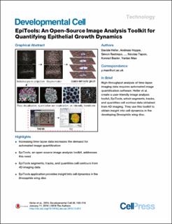
| dc.description.abstract | Epithelia grow and undergo extensive rearrangements to achieve their final size and shape. Imaging the dynamics of tissue growth and morphogenesis is now possible with advances in time-lapse microscopy, but a true understanding of their complexities is limited by automated image analysis tools to extract quantitative data. To overcome such limitations, we have designed a new open-source image analysis toolkit called EpiTools. It provides user-friendly graphical user interfaces for accurately segmenting and tracking the contours of cell membrane signals obtained from 4D confocal imaging. It is designed for a broad audience, especially biologists with no computer-science background. Quantitative data extraction is integrated into a larger bioimaging platform, Icy, to increase the visibility and usability of our tools. We demonstrate the usefulness of EpiTools by analyzing Drosophila wing imaginal disc growth, revealing previously overlooked properties of this dynamic tissue, such as the patterns of cellular rearrangements. | de_CH |
| dc.language.iso | en | de_CH |
| dc.publisher | Cell Press | de_CH |
| dc.relation.ispartof | Developmental Cell | de_CH |
| dc.rights | http://creativecommons.org/licenses/by/4.0/ | de_CH |
| dc.subject | Epithelial dynamics | de_CH |
| dc.subject | Image analysis | de_CH |
| dc.subject | Network description | de_CH |
| dc.subject | Cell Shape | de_CH |
| dc.subject | Epithelium | de_CH |
| dc.subject | Fluorescence microscopy | de_CH |
| dc.subject | Morphogenesis | de_CH |
| dc.subject | Cell tracking | de_CH |
| dc.subject | Computer-assisted image processing | de_CH |
| dc.subject.ddc | 005: Computerprogrammierung, Programme und Daten | de_CH |
| dc.subject.ddc | 571: Physiologie und verwandte Themen | de_CH |
| dc.title | EpiTools : an open-source image analysis toolkit for quantifying epithelial growth dynamics | de_CH |
| dc.type | Beitrag in wissenschaftlicher Zeitschrift | de_CH |
| dcterms.type | Text | de_CH |
| zhaw.departement | Life Sciences und Facility Management | de_CH |
| zhaw.organisationalunit | Institut für Computational Life Sciences (ICLS) | de_CH |
| dc.identifier.doi | 10.21256/zhaw-4376 | - |
| dc.identifier.doi | 10.1016/j.devcel.2015.12.012 | de_CH |
| dc.identifier.pmid | 26766446 | de_CH |
| zhaw.funding.eu | No | de_CH |
| zhaw.issue | 1 | de_CH |
| zhaw.originated.zhaw | Yes | de_CH |
| zhaw.pages.end | 116 | de_CH |
| zhaw.pages.start | 103 | de_CH |
| zhaw.publication.status | publishedVersion | de_CH |
| zhaw.volume | 36 | de_CH |
| zhaw.publication.review | Peer review (Publikation) | de_CH |
| Appears in collections: | Publikationen Life Sciences und Facility Management | |
Files in This Item:
| File | Description | Size | Format | |
|---|---|---|---|---|
| 2016_Heller_EpiTools_Developmental_Cell_36_1.pdf | 7.4 MB | Adobe PDF |  View/Open |
Show simple item record
Heller, D., Hoppe, A., Restrepo, S., Gatti, L., Tournier, A. L., Tapon, N., Basler, K., & Mao, Y. (2016). EpiTools : an open-source image analysis toolkit for quantifying epithelial growth dynamics. Developmental Cell, 36(1), 103–116. https://doi.org/10.21256/zhaw-4376
Heller, D. et al. (2016) ‘EpiTools : an open-source image analysis toolkit for quantifying epithelial growth dynamics’, Developmental Cell, 36(1), pp. 103–116. Available at: https://doi.org/10.21256/zhaw-4376.
D. Heller et al., “EpiTools : an open-source image analysis toolkit for quantifying epithelial growth dynamics,” Developmental Cell, vol. 36, no. 1, pp. 103–116, 2016, doi: 10.21256/zhaw-4376.
HELLER, Davide, Andreas HOPPE, Simon RESTREPO, Lorenzo GATTI, Alexander L. TOURNIER, Nicolas TAPON, Konrad BASLER und Yanlan MAO, 2016. EpiTools : an open-source image analysis toolkit for quantifying epithelial growth dynamics. Developmental Cell. 2016. Bd. 36, Nr. 1, S. 103–116. DOI 10.21256/zhaw-4376
Heller, Davide, Andreas Hoppe, Simon Restrepo, Lorenzo Gatti, Alexander L. Tournier, Nicolas Tapon, Konrad Basler, and Yanlan Mao. 2016. “EpiTools : An Open-Source Image Analysis Toolkit for Quantifying Epithelial Growth Dynamics.” Developmental Cell 36 (1): 103–16. https://doi.org/10.21256/zhaw-4376.
Heller, Davide, et al. “EpiTools : An Open-Source Image Analysis Toolkit for Quantifying Epithelial Growth Dynamics.” Developmental Cell, vol. 36, no. 1, 2016, pp. 103–16, https://doi.org/10.21256/zhaw-4376.
Items in DSpace are protected by copyright, with all rights reserved, unless otherwise indicated.